MICROFABRICATION
Chemical & Engineering News
August 20, 2001
Volume 79, Number 34
CENEAR 79 34 p. 14
ISSN 0009-2347
Working Microdevices Edge Closer To Reality
SOPHIE WILKINSON
"Fantastic Voyage"--the film in which a miniaturized Raquel
Welch and her colleagues venture through a patient's
bloodstream in a tiny submarine--no longer seems so
fantastical. Recent news reports have described a
camera-containing pill that photographs the digestive tract.
And Japanese researchers have now made microdevices
that could proceed through the body "through even the
smallest blood vessels, for example, to deliver clinical
treatments" [Nature, 412, 697 (2001)].
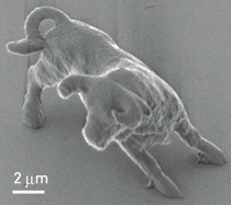
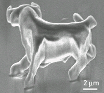
Applied physics professor Satoshi Kawata and coworkers at Osaka
University have crafted what they say are the smallest model animals
and
among the smallest functional micromechanical systems ever made. Their
"micro-bulls" are 10 µm long and 7 µm high, about the size
of a red blood
cell (see pictures below). Their similarly sized "micro-oscillator
system"
consists of a bead fastened to a spring attached to a cubic anchor.
The
scientists employ laser-trapping force to catch hold of the bead and
pull
on it. When released, the bead moves as the spring contracts and relaxes.
The Japanese team uses "two-photon photopolymerization" to create the
3-D
structures. An infrared laser is beamed into a liquid urethane-acrylate
resin
containing photoinitiators, and the resin solidifies wherever two photons
are
simultaneously absorbed. Movement of the laser's focal point location
is
managed by computer. After the pattern is completed, unreacted resin
is
washed away. The researchers bettered the technique's previous minimum
feature size of 600 nm by controlling laser-pulse energy and exposure
time to
give a resolution of 120 nm.
DOWNSIZED The fine features of the bull and the functionality of the
ball-on-a-spring device demonstrate the laser technique's capabilities.
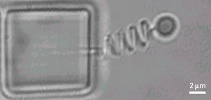
©NATURE