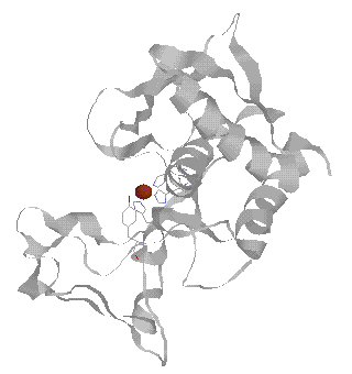
Crayfish astacin is a unique small Zn2+ endopeptidase (MW = 22 kDa) found in the digestive fluid of crustaceans, and belongs to the only Zn protein family that contain a coordinated Tyr under physiological conditions [1] (see structure). However, the presence of a negatively charged phenolate, i.e., the coordinated Tyr, is known to decrease the Lewis acidity of the metal ion in metal complexes [2,3]. As a result, a decrease in water activation by ~2 orders in terms of the pKa of the coordinated water has been observed in metal complexes. This decrease in Lewis acidity of the metal by a coordinated phenolate should presumably result in a significant decrease in metal-centered hydrolytic activity. On the contrary, this is not the case in phenolate(Tyr)-coordinated astacin which still exhibits high hydrolytic activity under neutral conditions. Hence, the coordinated Tyr may not act to lower the Lewis acidity of the active-site metal in astacin during its catalysis, but may play some unique roles in the action of astacin that have not yet been observed in other metalloproteases. A better understanding of the roles of this coordinated Tyr is essential to provide further insight into the catalysis of astacin family.
Several zinc proteins [4-6], such as bone morphogenetic protein-1, hydra metalloprotease 1, the embryonic hatching proteins from medakafish and sea urchin, and the alkaline endopeptidases from Serratia and Pseudomonas species have been revealed by means of sequence alignment to contain conserved metal binding residues as in astacin, i.e., 3 His residues found in the consensus sequence HEXXHXXGXXH and 1 Tyr in a loop containing Met-His(or Ser)-Tyr. The three-His sequence is also found to be the consensus sequence in snake venom proteases and collagenases [4-6]. Since astacin is the simplest member in this Zn protease family, it may serve as a prototype which may offer us the opportunity to study and understand the mechanism of this new protease family.
Despite the high Lewis acidity of Cu2+ ion [2,3,7], almost all Cu2+-substituted derivatives of Zn2+ hydrolytic enzymes are inactive [7]. A few Cu2+-substituted derivatives of Aeromonas aminopeptidase were determined to exhibit super-activities compared with the native enzyme toward some poor substrates, although these activities are still far below the activity of the native enzyme toward the most efficient substrate [8]. Cu2+-substituted astacin exhibits surprisingly high activity against the tripeptide substrate succinyl-tri-Ala-nitroanalide (37% that of the native enzyme in terms of kcat/Km [9]), thus deserves a thorough study to shed light on this unique Cu2+ activation of this hydrolytic enzyme. Conversely, full activities are usually observed in the case of Co2+-substituted derivatives of Zn enzymes, such as astacin [9], and are occasionally higher than those of the native enzymes [7,9]. Due to the presence of unpaired electrons in Cu2+ and Co2+ ions, various spectroscopic and magnetic techniques can be applied to the study of their binding environments in proteins. Thus, Cu2+- and Co2+-substituted derivatives of astacin can serve as very good model systems for the study of astacin action by means of spectroscopic and magnetic techniques.
We have been studying crayfish astacin and Serratia metalloprotease (serralysin) using Co2+ and Cu2+ as spectroscopic and magnetic probes. Our studies have shown that the coordinated Tyr in these proteins are detached upon inhibitor binding, which may suggest the same binding status during catalysis (see abstract). Our current studies on this protein family include a complete kinetic studies of the metal derivatives of serralysin, pulsed EPR studies of the Cu2+ derivatives of serralysin and astacin in collaboration with Dr. Alexander Angerhofer at University of Florida, and detailed NMR analysis of Co2+-astacin and its inhibitor complexes.
References
[1] W. Stöcker and R. Zwilling, Met.
Enzymol. 248, 305 (1995).
[2] E. Kimura, Prog. Inorg. Chem. 41,
443 (1994).
[3] E. Kimura and T. Koike, Adv. Inorg. Chem.
44, 229 (1997)
[4] W. Stöcker, F. Grams, U. Baumann,
P. Reinemer, F.-X. Gomis-Rüth,D. B. McKay and W. Bode, Protein
Sci. 4, 823
(1995).
[5] J. S. Bond and R. J. Beynon, Protein Sci.
4, 1247 (1995).
[6] M. P. Sarras, Jr., BioEssays 18,
439 (1996).
[7] I. Bertini and C. Luchinat in Bioinorganic
Chemistry, I. Bertini, H. B. Gray, S. J. Lipard and J. S. Valentine,
Eds.,
University Science
Books, CA, 1994, Chapter 2.
[8] M. E. Bayliss and J. M. Prescott, Biochemistry
25, 8113 (1986) and reference therein.
[9] F.-X. Gomis-Rüth, F. Grams, I. Yiallouros,
H. Nar, U. Küsthardt, R. Zwilling, W. Bode and W. Stöcker, J.
Biol. Chem.
269, 17111
(1994).